Role of Imaging and Biopsy in Bone Tumour Diagnosis

Introduction to Bone Tumour Diagnosis

Accurate diagnosis is the cornerstone of successful bone tumour treatment. Bone tumours may be benign or malignant, and distinguishing between them requires a structured and careful diagnostic process. Imaging and biopsy play a vital role in bone tumour diagnosis by helping doctors identify the nature, location, and aggressiveness of the tumour. For patients seeking care from a Bone Cancer Specialist in Dehradun, understanding why multiple investigations are required can ease anxiety and improve confidence in treatment decisions.

Why Accurate Bone Tumour Diagnosis Is Essential

Bone tumours often mimic common orthopaedic problems such as sports injuries, arthritis, or infections. Delayed or incorrect diagnosis can lead to disease progression and limited treatment options. Proper bone tumour diagnosis ensures timely intervention, avoids unnecessary surgeries, and improves outcomes. Imaging and biopsy together form the foundation of reliable diagnosis and guide safe, effective treatment planning.

Clinical Evaluation and the Role of Imaging in Bone Tumour Diagnosis

The diagnostic journey begins with a detailed clinical evaluation, including medical history, pain characteristics, swelling, and functional limitations. Based on these findings, imaging becomes the first major step in bone tumour diagnosis.

X-ray, MRI, and CT Scan in Bone Tumour Evaluation

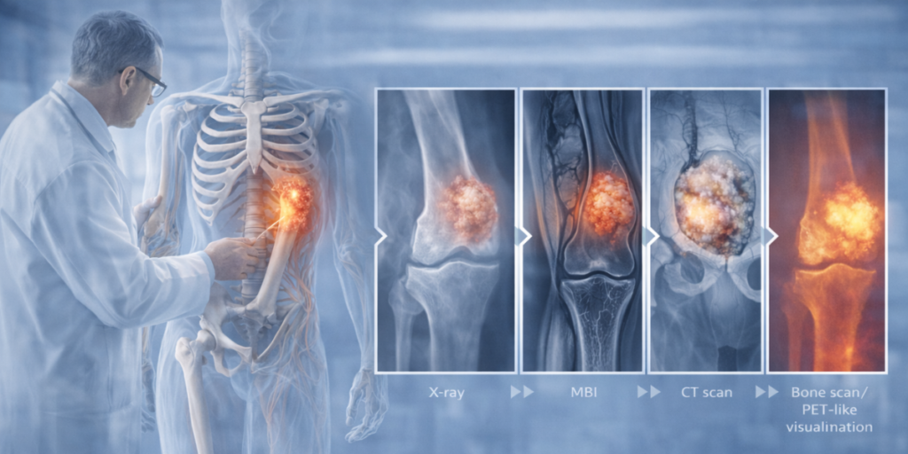
X-rays are usually the initial imaging modality, offering valuable information about bone destruction, abnormal growth, and tumour location. Certain patterns on X-rays can suggest whether a lesion is aggressive or benign.

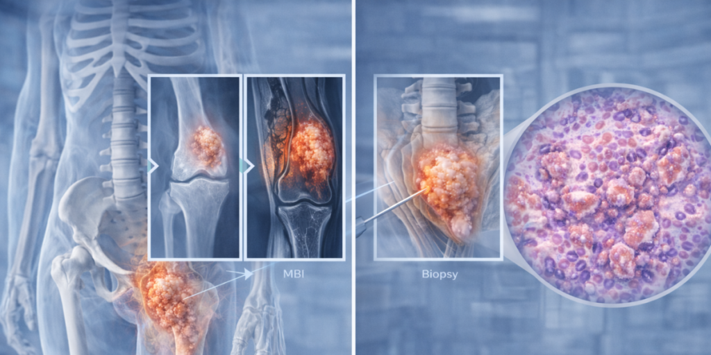
MRI plays a crucial role in evaluating soft tissue involvement and tumour extent. It helps determine whether surrounding muscles, nerves, or blood vessels are affected, which is essential for limb preservation and surgical planning. CT scans provide detailed visualization of bone structure and are especially useful in complex areas such as the pelvis and spine.

For patients consulting a Bone Cancer Specialist in Dehradun, access to advanced MRI and CT imaging allows for precise assessment and safe treatment planning.

Bone Scan and PET Scan for Staging

Bone scans and PET scans are used to detect tumour spread and identify multiple bone lesions. These imaging tools help determine whether the tumour is localized or metastatic, which directly influences treatment strategy and prognosis. Imaging features such as rapid bone destruction, irregular margins, and associated soft tissue mass often raise suspicion for malignant bone tumours and indicate the need for urgent biopsy.

Role of Biopsy in Bone Tumour Diagnosis

While imaging strongly suggests the nature of a bone tumour, biopsy provides definitive confirmation. A biopsy determines whether the tumour is benign or malignant and identifies the exact tumour type. Treatment should never begin without histological confirmation, making biopsy a critical step in bone tumour diagnosis.

Biopsy Techniques and Proper Planning

Biopsy can be performed using needle techniques or open surgical methods, depending on tumour location and imaging findings. Proper planning is essential, as an incorrectly placed biopsy can complicate future surgery or increase the risk of tumour spread. For this reason, biopsy should always be performed by or in consultation with an experienced Bone Cancer Specialist in Dehradun who understands tumour anatomy and surgical planning.

Correlation Between Imaging and Biopsy Findings

Imaging and biopsy are complementary. Imaging guides the biopsy site, ensuring representative tissue sampling, while biopsy confirms imaging-based suspicions. This correlation allows accurate tumour classification, staging, and personalized treatment planning. Multidisciplinary collaboration between radiologists, pathologists, and orthopaedic oncologists further improves diagnostic accuracy and outcomes.

Common Pitfalls and the Importance of Specialist Care

One of the most common mistakes in bone tumour diagnosis is performing surgery or biopsy without adequate imaging or specialist input. Early referral to a Bone Cancer Specialist in Dehradun helps avoid diagnostic errors, ensures safe biopsy, and improves long-term results.

Advanced Bone Tumour Diagnostic Facilities in Dehradun

Dehradun now offers comprehensive diagnostic services for bone tumour evaluation. Patients have access to advanced imaging and biopsy facilities under expert supervision, enabling timely and accurate diagnosis close to home.

Diagnostic Facilities Available

  • Advanced X-ray, MRI, and CT imaging
  • Imaging-guided biopsy procedures
  • Accurate histopathological diagnosis
  • Multidisciplinary tumour board evaluation
  • Personalized treatment planning

About Dr. Narendra Singh Butola – Bone Cancer Specialist in Dehradun

Dr. Narendra Singh Butola is a highly experienced Orthopaedic Oncologist and Sarcoma Specialist, recognized as the first bone cancer specialist in Uttarakhand. He specializes in the accurate diagnosis and treatment of benign and malignant bone and soft tissue tumours.

Professional Profile
Name: Dr. Narendra Singh Butola
Designation: Orthopaedic Oncologist & Sarcoma Specialist
Expertise:

  • Diagnosis of benign and malignant bone tumours
  • Imaging-guided biopsy planning
  • Limb salvage surgery

Hospital: Graphic Era Institute of Medical Sciences
Location: Chakrata Road, Dhulkot, Dehradun, Uttarakhand – 248007, India

Conclusion

The role of imaging and biopsy in bone tumour diagnosis is fundamental. Imaging provides crucial information about tumour location, extent, and aggressiveness, while biopsy confirms the exact diagnosis. Together, they form the backbone of effective treatment planning. Early evaluation by a Bone Cancer Specialist in Dehradun ensures safe biopsy, accurate diagnosis, and better outcomes. With advanced diagnostic facilities at Graphic Era Institute of Medical Sciences and expert care under Dr. Narendra Singh Butola, patients receive precise, compassionate, and timely bone tumour diagnosis—laying the foundation for successful treatment and recovery.